Green sea turtles are listed as threatened or endangered. Credit: Thierry Work, USGS.
A group of scientists including Tina Weatherby of University of Hawai‘i at Mānoaʻs School of Ocean and Earth Science and Technology (SOEST) have for the first time successfully grown reptile skin in a laboratory in an effort to better understand how to treat a viral disease affecting green sea turtles.
The international effort, led by the U.S. Geological Survey, engineered turtle skin to grow a virus called chelonid herpesvirus 5 or ChHV5, a tumor disease associated with fibropapillomatosis (FP) which affects sea turtles worldwide, but particularly those in Hawai‘i, Florida and Brazil. FP causes disfiguring tumors on the eyes, skin and mouth of green turtles, as well as internal tumors. The virus also compromises the reptileʻs immune system, leaving it vulnerable to infection and emaciation which is often fatal.
The studyʻs results bring researchers a step closer to understanding and fighting the viral disease that threatens the already endangered species.
ARTICLE CONTINUES BELOW AD
ARTICLE CONTINUES BELOW AD
“Fibropapillomatosis is the most common infectious disease affecting endangered green turtles,” said USGS Scientist Thierry Work, lead author of the study. “Our findings provide a significant advancement in studying FP, and may eventually help scientists better understand other herpes virus-induced tumor diseases, including those of humans.”
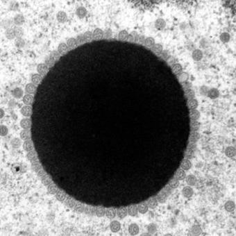
TEM image shows a sun-shaped area in turtle cells where ChHV5 replicates. Credit: Thierry Work, USGS.
Using cells from both tumors and normal turtle skin, scientists reconstructed the complex structure of turtle skin, enabling the growth of ChHV5 in the lab. Weatherby of SOEST cut precise, ultrathin slices of the skin at roughly 60 to 80 nanometers in thickness–about one thousandth the diameter of a strand of hair–to observe the virus replicating in unprecedented detail using a transmission electron microscope.
ChHV5 was discovered more than 20 years ago, but little was understood about how it causes tumors due to a lack in blood tests and the previous inability to grow it in a laboratory.
ARTICLE CONTINUES BELOW AD
“Examining viruses within the complex three-dimensional structure of engineered skin is exciting, because virus replication in such a system is likely much closer to reality than traditional laboratory techniques,” Work said. “This method could be a powerful tool for answering broader questions about virus-induced tumors in reptiles and herpes virus replication in general.”
The USGS partnered with the University of Hawai‘i, the National Oceanic and Atmospheric Administration and the University of Zurich to conduct the study.
This comments section is a public community forum for the purpose of free expression. Although Big Island Now encourages respectful communication only, some content may be considered offensive. Please view at your own discretion. View Comments